Optimizing Imaging Protocols and Software for Live Animal Imaging
Award Imaging Scientist
Funding Cycle Cycle 1
Investigator

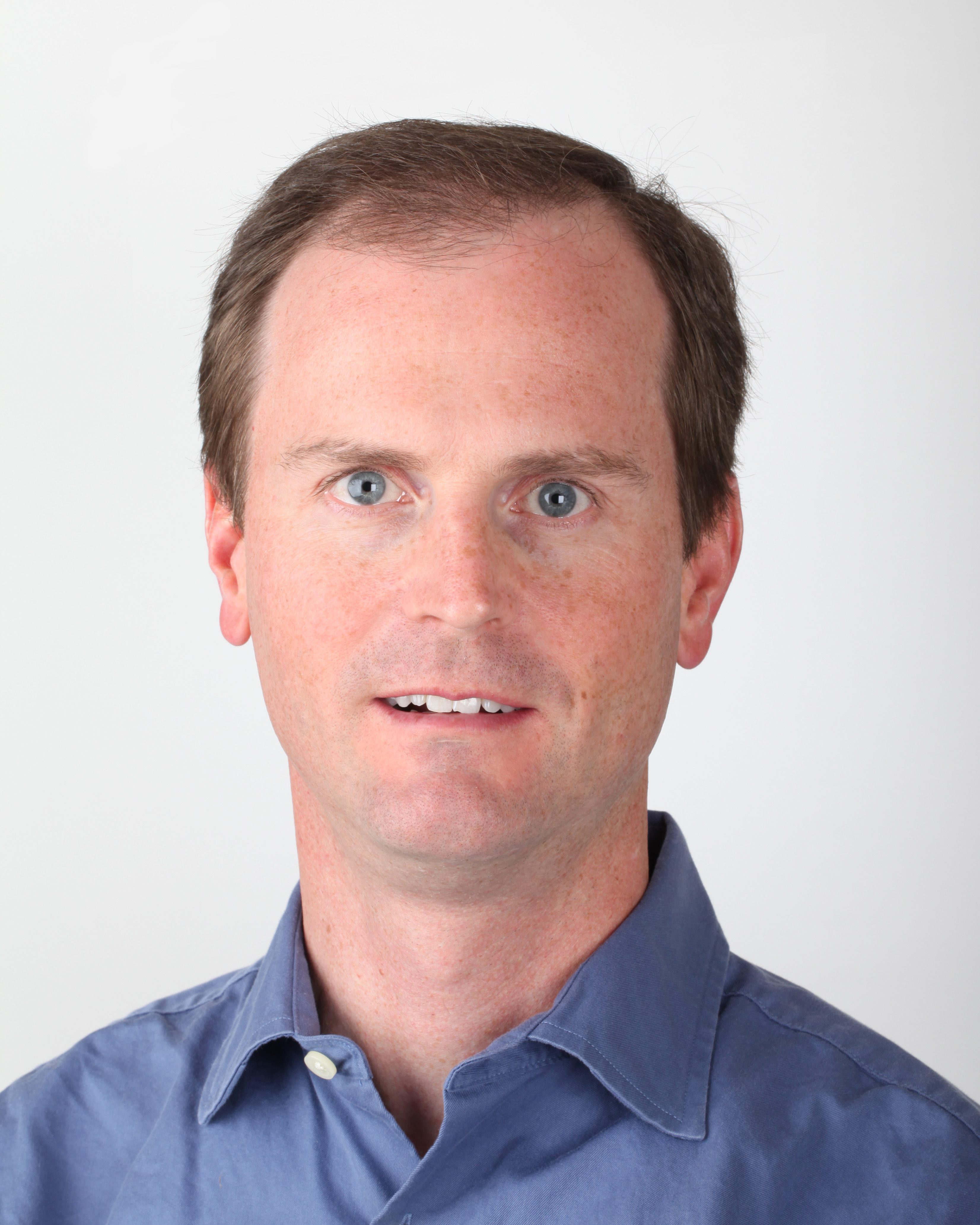
Douglas J. Rowland, PhD
University of California, Davis (Center for Molecular and Genomic Imaging)
Bio
Dr. Rowland has over 18 years of experience in biomedical imaging and six years in instrumentation for physics experiments. His specific focus was on the production, chemistry and characterization of non-standard PET radionuclides in the first years of work in biomedical imaging. From there, he transitioned to developing techniques for imaging small animals on microPET and microCT technologies. Since 2007, he has been responsible for collaborating with principal investigators on the design and implementation of imaging experiments on small animal in vivo imaging technologies, including MRI and PET. He also worked to establish a strong MRI and CT research program at CMGI, particularly in high resolution imaging for correlative microscopy.
Project Description
Dr. Rowland works with instruments that allow for live animal imaging, similar to how people are scanned in the clinic, such as MRI, CT or PET scanners. These preclinical technologies are used to understand animal models for human disease. Scanners in CMGI, such as the CT and MRI scanners, are capable of assessing tissues down to the level of a few microns to tens of millimeters. Dr. Rowland will refine and optimize imaging protocols and software for these scanners to correlate with microscopy. He will also consult with scientists to identify their image analysis bottlenecks and needs, and work collaboratively to document unsolved image analysis challenges and communicate them to the wider imaging communities. Additionally, he will create instructional videos to cover basic and advanced image analysis approaches.


