
Oct 17, 2024 · 6 min read
Nobel Laureate David Baker, Colleagues Design Proteins for Advanced Microscopy
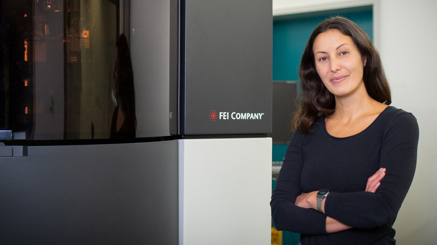
David Baker’s 2024 Nobel Prize-winning work is being used in collaboration with Julia Mahamid to design new self-assembling protein labels for electron microscopy.

Try to imagine creating a new color that has never existed before. Biochemist Dr. David Baker was awarded one-half of the Nobel Prize in Chemistry 2024 for achieving something similar — not for colors, but for proteins. By designing new proteins from the 20 naturally occurring amino acids, he and his team have created molecules that never existed in nature, with potential uses in pharmaceuticals, vaccines, nanomaterials and sensors.
A single human cell can contain billions of proteins, the building blocks and core workers in cells, each driving virtually everything that makes cells function and allows them to assemble into tissues and organisms. Proteins help us see, move our muscles, catalyze the reactions that give us energy, serve as antibodies that help us fight infections, and act as hormones that signal key bodily functions. Understanding their structure and how they interact with one another and different parts of the cell is fundamental to our study of human biology.
This area of research is so fundamental to biology that it has been recognized with numerous Nobel Prizes. In 1972, Christian Anfinsen was awarded the Nobel Prize in Chemistry for discovering that the 3D structure of proteins is determined by their sequence of amino acids. This truth sparked an irresistible hope and journey of discovery for many scientists: if a protein structure, and thus its function, could be predicted based on its amino acid sequence, it would revolutionize our understanding of health and disease.
For decades, predicting protein structures remained one of biology’s grandest challenges. In the late 1990s, early work in the Baker lab led to the development of Rosetta. This computer program made major leaps in accurately predicting how proteins fold into their complex 3D structures. Groundbreaking on its own — and with modern adaptations still in use today — it’s what he did next that changed the course of his career and eventually led to the Nobel Prize.
The Big Breakthrough: Designing New Proteins From Scratch
Part of the draw to knowing protein structures is to harness their functions, whether it’s restoring malfunctioning proteins in a disease or improving enzymatic digestion of harmful substances. But instead of being limited to the repertoire of proteins that already exist, Baker realized they could design their own proteins with desirable functions from scratch, then use the software to figure out the amino acid sequence that could make this entirely new protein possible.
Baker achieved this massive breakthrough in a paper published in 2003 with an entirely new protein, unlike anything existing in nature, called Top7. This feat skyrocketed the field of protein design, making the dream of newly created proteins as chemical tools a reality.
“David Baker’s work has almost single-handedly driven the field of protein design,” says biochemist David Agard, founding scientific director at the Chan Zuckerberg Imaging Institute and head of the lab at the University of California San Francisco (UCSF) where Baker completed his postdoctoral research.
Baker is exactly the right person to be recognized by the Nobel Prize.
Since then, Baker’s lab has been applying this technology to design proteins that solve challenges in medicine. This includes designing new proteins that can function as pharmaceuticals to more precisely treat disease, and proteins that can act as new biosensors to study the interplay between cancer, cancer treatments and cell signaling. Before the 2024 Nobel, Baker received a myriad of accolades for this work, including the 2021 Breakthrough Prize in Life Sciences.
Agard adds, “Protein design has the potential not only to revolutionize clinical therapeutics, but to be transformative in its ability to create custom eco-friendly nanomaterials that will have broad societal impact. Baker is exactly the right person to be recognized by the Nobel Prize.”
What’s Next After Winning a Nobel?
One of Baker’s current projects is to design proteins that can be used to help reveal the inner workings of cells at atomic resolution. As a grantee in CZI’s Frontiers of Imaging Technology program, Baker is collaborating with imaging expert Dr. Julia Mahamid to create a universal label for an advanced imaging technique called cryo-electron tomography (cryoET).
CryoET is revolutionary for understanding biology because it uniquely enables researchers to visualize the structure of proteins in atomic-level detail — while maintaining the overall cellular architecture of the sample. This gives researchers a close-up view of proteins in their original biological context to pinpoint what a healthy cell looks like and what changes may occur in disease.
The density and complexity of the cell, however, make finding a protein (or group of proteins) of interest, like searching for a needle in a haystack. The collaboration leverages Mahamid’s world-renowned expertise in cryoET and Baker’s leadership in protein design to shine a beacon on the needle. To do this, they’re designing proteins that can self-assemble into unique shapes that make them detectable in cryoET. These self-assembling proteins will serve as labels to locate targets within the cell for analysis at atomic resolution.
There has been a longstanding need for a genetic label that would act as a universal beacon in cryoET, just as green fluorescent protein (GFP) is for fluorescence microscopy. The discovery of GFP opened a whole new window into visualizing biological processes within our cells using light microscopy. In fact, Osamu Shimomura, Martin Chalfie and Roger Y. Tsien were awarded the Nobel Prize in Chemistry in 2008 for the countless processes within the cell GFP revealed through light microscopy. However, this type of labeling technique is incompatible with electron microscopy, so developing a universal marker that works simultaneously in fluorescence microscopy and cryoET could similarly propel the field of molecular cell biology forward.
“Their work is critical to the field of cryoET,” Agard says. “It will combat a long-standing major bottleneck in studying the structure of biological molecules in their natural environment.”
Advanced imaging gives researchers a detailed view of the mechanisms underlying health and disease and — together, with the open sharing through the CryoET Data Portal and CZI’s broader work to understand the mysteries of the cell and how cells interact within systems — can ultimately drive diagnostics and inform new treatments.
Given the potential of Baker and Mahamid’s new technology, perhaps there might be another call from Sweden in the future.
Learn more about our Frontiers of Imaging Technology grantees and the groundbreaking research we’re doing at the Chan Zuckerberg Institute for Advanced Biological Imaging.


Sorry, marketing cookies are required to view this form.