
Oct 11, 2023 · 6 min read
Building Local Solutions for Accessing Imaging Technology
This local leader is committed to finding solutions for improving accessibility to powerful imaging technology in South America.
Imaging technologies continue to propel biomedical discoveries forward with advancements in new super-resolution microscopy techniques, analysis algorithms and live-imaging methodologies, to name a few. However, the availability of new technologies does not equate to accessibility, and expertise and access are limited in many countries due to ongoing socioeconomic, educational and technological challenges.
Global access to imaging technologies is critical for a more just and healthy future. It enables researchers to pursue research that is a high priority for their local context, ensuring global representation in research. Further, it strengthens all biomedical research to have a diversity of expertise and feedback, as well as applications of research.
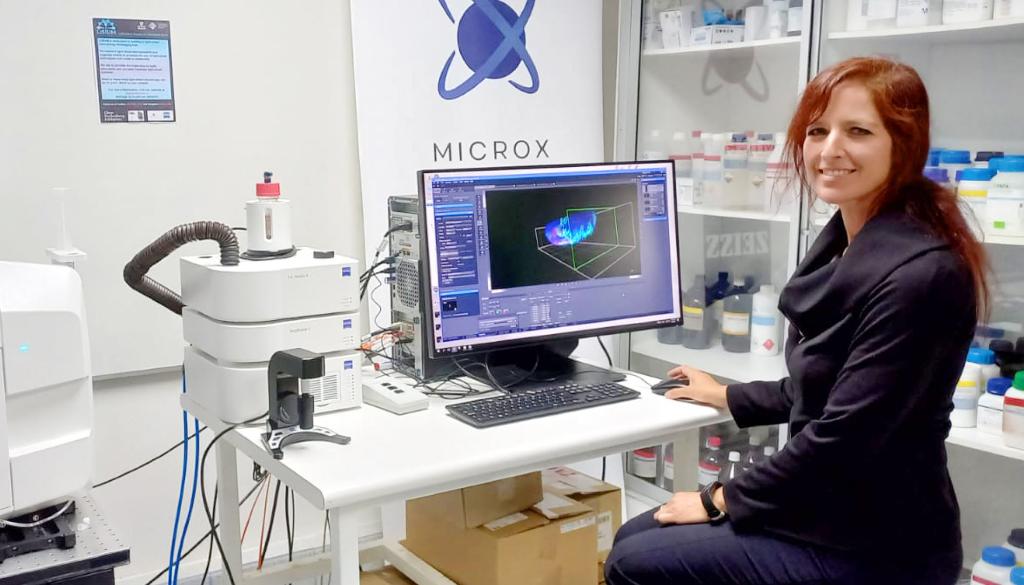
Dr. Alenka Lovy, an assistant professor at the Universidad Mayor, is building capacity for light-sheet imaging across labs in her university, the rest of Chile and South America. She leads a network of scientists building tailored solutions to connect more researchers in the region with both commercial and portable light-sheet microscopes to enhance their research.
Lovy’s Love of Light-Sheet
Lovy’s current research focuses on how certain bacteria can affect aging and mitochondrial function. As a cell biologist by training, Lovy was no stranger to spending hours at the microscope. But when her institute acquired a commercial light-sheet microscope in 2021, it was the first on the continent and the first time she and many other researchers had a chance to work with one.
I find it very exciting that we can collect live movies of biological processes with the light-sheet microscope that we couldn’t do with the confocal.
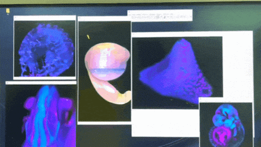
Light-sheet microscopy is a powerful tool for 3D imaging of intact living model organisms. It uses a thin sheet of light to excite just the fluorophores within the focal area. This allows them to capture optical sections, including axial resolution (the ability to discern two structures along or parallel to the path of the fluorescent beam).
Because of this setup, light-sheet microscopy has the advantage of limiting the area of the sample subject to photobleaching and phototoxicity, meaning sample integrity remains over longer imaging periods. Light-sheet imaging can also record tens to thousands of images per second. It’s ideal for detailed analyses of large, optically cleared samples, providing high spatio-temporal resolution for live-imaging biological specimens over longer periods.
Lovy was eager to try the light-sheet microscope at the Universidad Mayor for her research.
“I was really excited to start to learn more about this technology,” she says. “But our biggest mission is to share the technology with others because it’s the only one of its kind basically on the continent.”
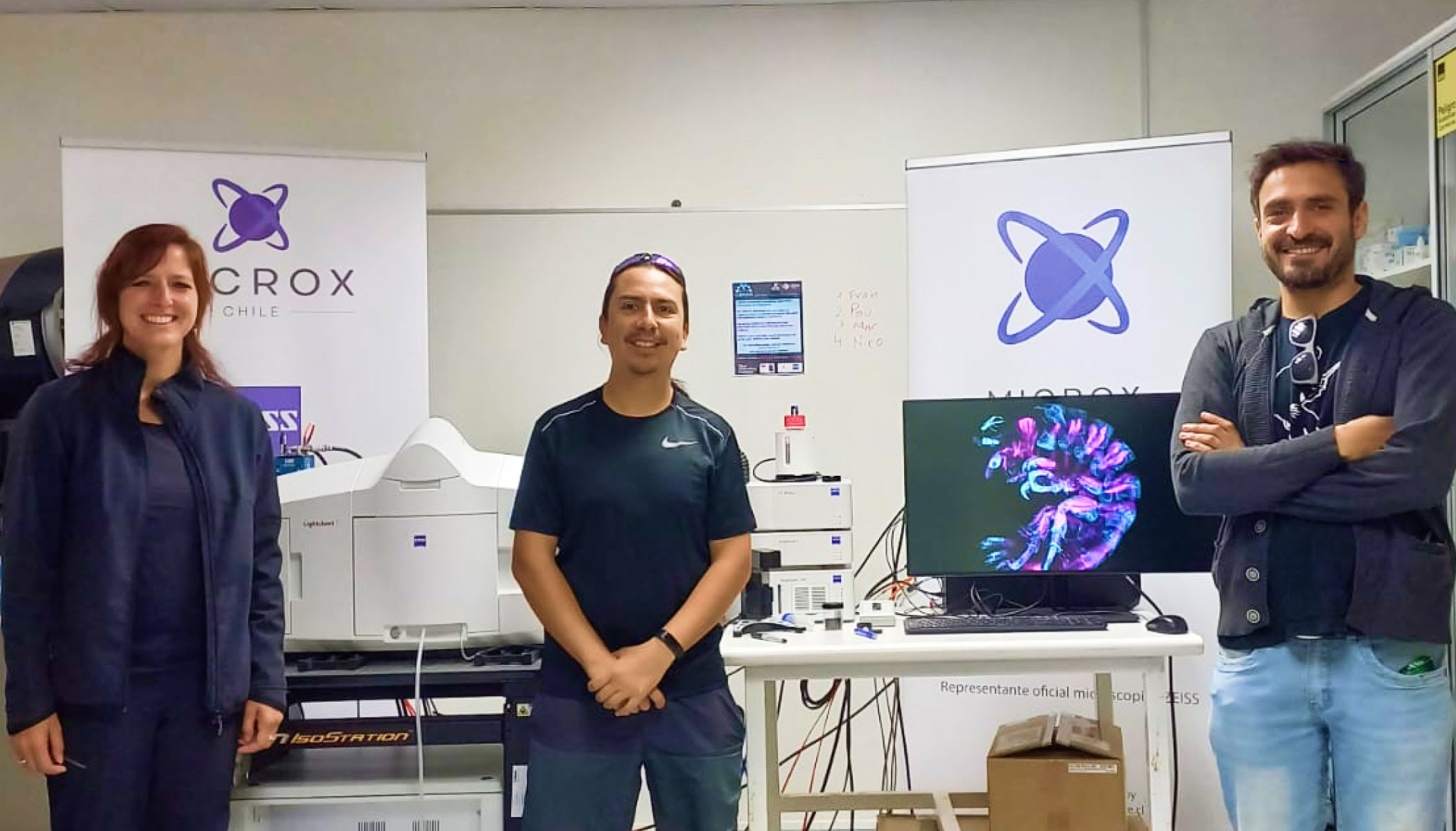
To enable this, Lovy led an Expanding Global Access to Bioimaging grant application alongside Felipe Court and Leonardo Valdivia to begin building networks of researchers called the Light-Sheet Imaging at Universidad Mayor (LiSIUM). LiSIUM connects five labs at Universidad Mayor, five labs in other institutions in Chile, and four international labs in South America to share information about this instrument.
The hub also leverages workshops, conferences and experts to make the technology more accessible for biomedical researchers. For example, Lovy’s team coordinated training opportunities offered concurrently with a world-class European Molecular Biology Organization developmental biology course, bringing together experts in model organisms suitable for light-sheet imaging.
Building Sustainable for Sustainable Access
Once Lovy’s team established a foundational network for light-sheet microscopy, she wanted to find a way to remove additional barriers to accessing this technology. When attending a light-sheet microscopy conference in 2020, Lovy learned about the compact and portable light-sheet microscope developed by Jan Huisken, nicknamed “Flamingo” for its one-legged stand and vertical profile. Not only is the Flamingo light-sheet microscope small enough to fit into a suitcase, but it’s also relatively easy to set up in a new location with the remote configuration offered by Huisken as needed.
After contacting Huisken, Lovy and collaborators applied for an Advancing Imaging Through Collaborative Projects grant with collaborators at her university in Chile, Brazil, Uruguay and Germany to build three more affordable, portable light-sheet microscopes.
By collaborating with regional imaging centers, the new microscopes will be well integrated into biomedical centers that can use them.
“It’s really hard to come by this very expensive technology here,” Lovy shares, “and the idea of being able to travel with a microscope or send it to people that need it is a very exciting one.”
But as with all aspects of research, success is not reached overnight. The prohibitive cost and regulations surrounding importing parts mean they’re taking a combined approach of building locally and importing already-made instruments depending on the receiving country. Ultimately, the open sharing of a streamlined approach to building and operating these microscopes by Huisken means that once these logistics are optimized, more institutes can follow suit. Growing the imaging community in South America also means there will be more capacity to innovate other areas of imaging infrastructure, such as storage and data analysis, which currently present a challenge for newer imaging centers.
Read more: 6 Tips From Early Career Researchers To Remember on Good Days and Bad
For Lovy, it’s important to be transparent about administrative challenges related to this work because many researchers face them when trying to set up their labs or implement new technologies. These hurdles are just one of many challenges researchers navigate without a playbook, similar to the “hidden curriculum” many first-generation graduate students face. These challenges also underscore the difference between availability and accessibility, highlighting the importance of individual researchers leading the charge and modeling the changes needed for equitable global access in science.
Regional experts can solve challenges in a manner that makes sense for their local context, which is why the community that Lovy and her colleagues are building is so important.
Learn more about leaders making imaging technology and tools more accessible globally.


Sorry, marketing cookies are required to view this form.