To cure, prevent, and manage all diseases by the end of the century, we need a much deeper understanding of biological systems. Existing imaging tools provide a limited view, tending to focus on a specific biological scale without the necessary context. Researchers struggle to handle large volumes of data and to make quantitative insights, and substantial access and training gaps remain.
CZI’s imaging program aims to drive the development of:
A suite of new imaging tools capable of observing biological processes across spatial scales at the level of tissues, cells, and proteins; and
Robust frameworks to quantify, analyze, and share imaging data and share methods and tools.
Scientific imaging research and technology will play a critical role in enabling a deeper mechanistic understanding of health and disease, driving diagnostics, and informing directed treatments.
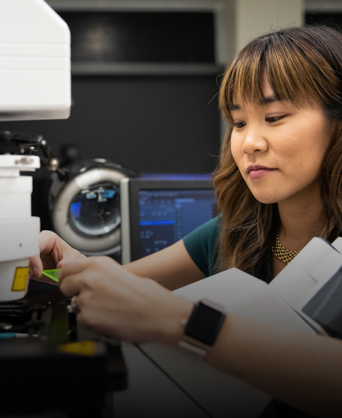
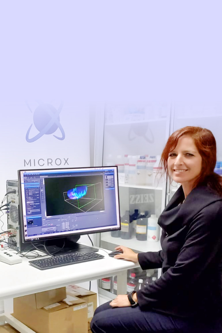
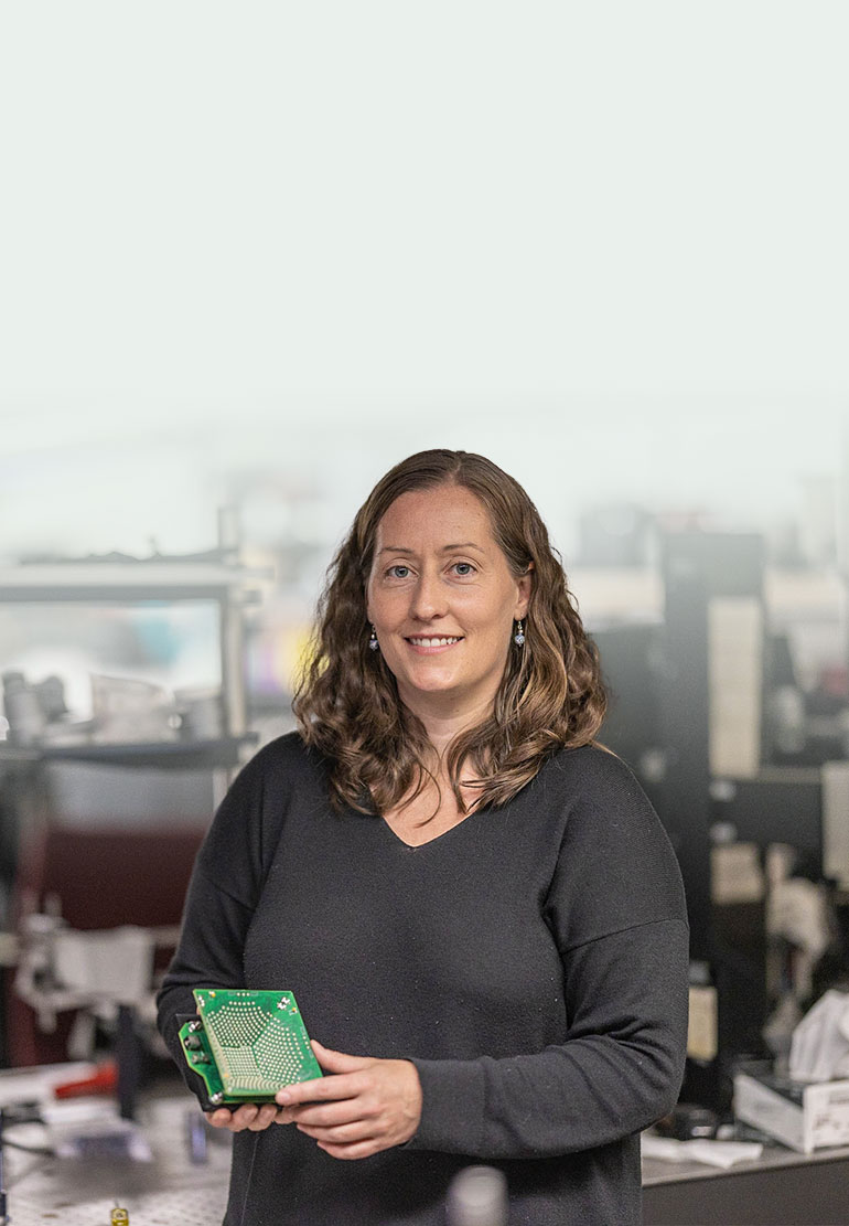
CZI Imaging Scientist Michelle S. Itano at the University of North Carolina, Chapel Hill, Neuroscience Microscopy Core. Photo provided by Michelle S. Itano.
CZI Imaging Scientist Michelle S. Itano at the University of North Carolina, Chapel Hill, Neuroscience Microscopy Core. Photo provided by Michelle S. Itano.
The Imaging program has funded the following RFAs:
Imaging Scientists (Cycle 1, $17.8 million and Cycle 2, $10.2 million)
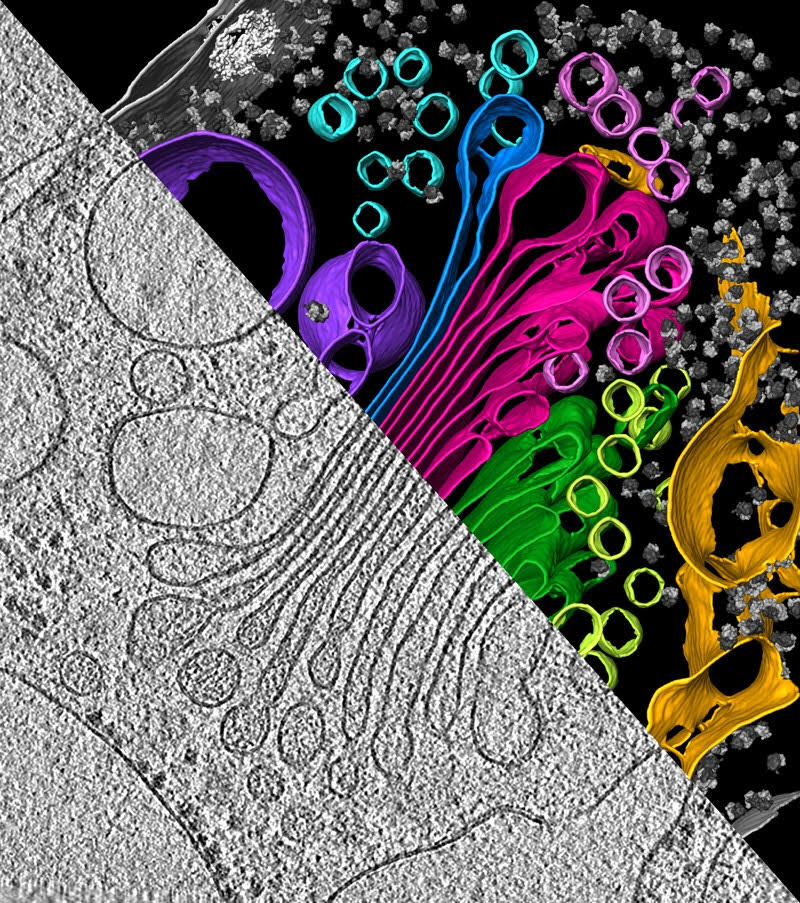
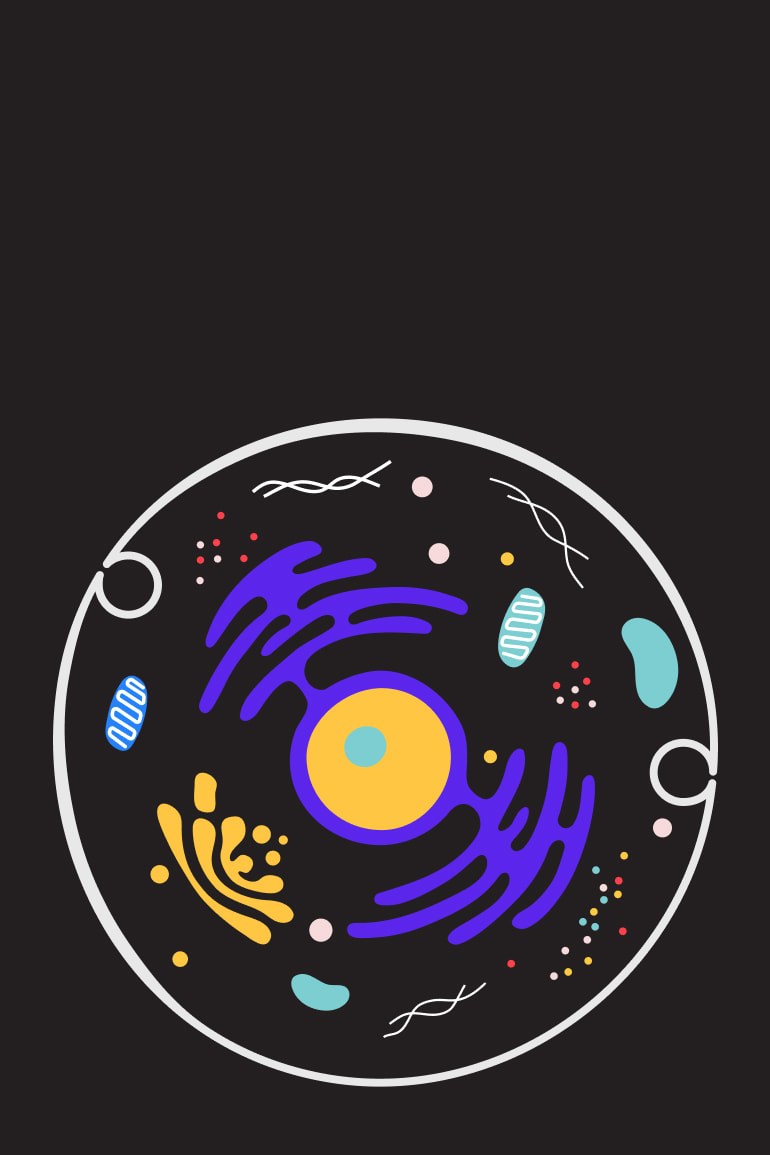
The inner workings of an algal cell as depicted with cryo-electron tomography, which aggregates multiple snapshots of a single piece of material. Visible are the endoplasmic reticulum (yellow), Golgi apparatus (green and magenta), and vesicles (multi-colored circles). | Photograph by Y. S. Bykkov et al./eLIFE (CC BY 4.0)
The inner workings of an algal cell as depicted with cryo-electron tomography, which aggregates multiple snapshots of a single piece of material. Visible are the endoplasmic reticulum (yellow), Golgi apparatus (green and magenta), and vesicles (multi-colored circles). | Photograph by Y. S. Bykkov et al./eLIFE (CC BY 4.0)
Chan Zuckerberg Institute for Advanced Biological Imaging
To cure, prevent, or manage all diseases by the end of the century, we need to develop a dynamic, integrated view of biological systems in health and disease. To accomplish this goal, the Chan Zuckerberg Initiative is creating a new advanced biological imaging institute. Located in California's Bay Area, disciplines will come together to develop revolutionary new imaging hardware and software tools that provide comprehensive views of biological systems in their native context.
CZI collaborates with napari — a community-built, Python-based, open source tool designed for browsing, annotating, and analyzing large multi-dimensional images. The napari hub is a CZI project, built and managed to support the napari community. This tool allows users to find community-built plugins that solve their analysis needs.
“We want napari to help not just Python practitioners, but also biologists and other scientists who want to access Python's enormous scientific ecosystem.”
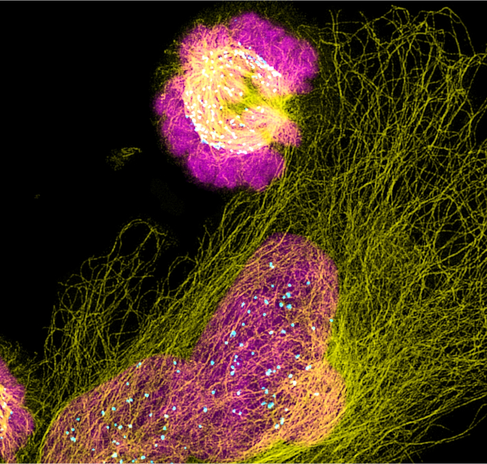
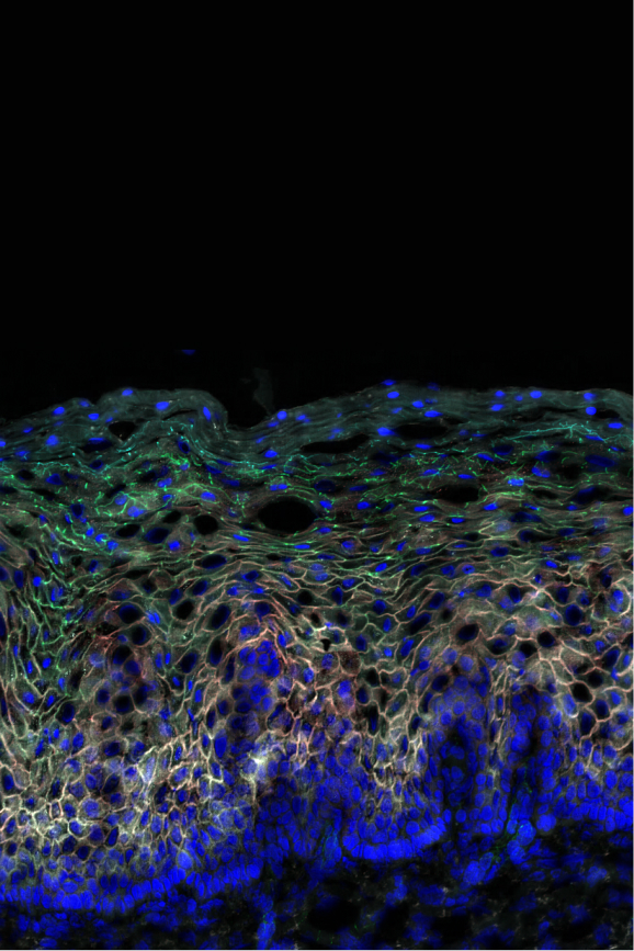
Super-resolution light microscopy allows scientists to view the details of subcellular organelles — units in cells with specialized functions — in living cells. Photo provided by Aaron Taylor, University of Michigan, BRCF Microscopy Core.
Super-resolution light microscopy allows scientists to view the details of subcellular organelles — units in cells with specialized functions — in living cells. Photo provided by Aaron Taylor, University of Michigan, BRCF Microscopy Core.
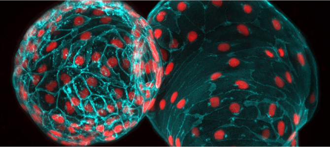
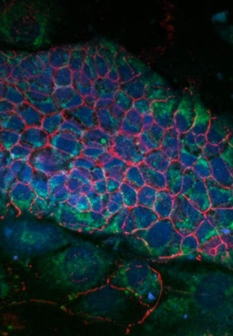
A two-day-old zebrafish heart viewed through a microscope. The heart muscle membrane is shown in blue and its nuclei in red. Photo provided by CZI imaging scientist Michael Weber of the Morgridge Institute for Research, in affiliation with the University of Wisconsin-Madison.
A two-day-old zebrafish heart viewed through a microscope. The heart muscle membrane is shown in blue and its nuclei in red. Photo provided by CZI imaging scientist Michael Weber of the Morgridge Institute for Research, in affiliation with the University of Wisconsin-Madison.
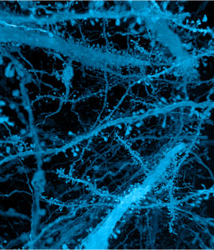
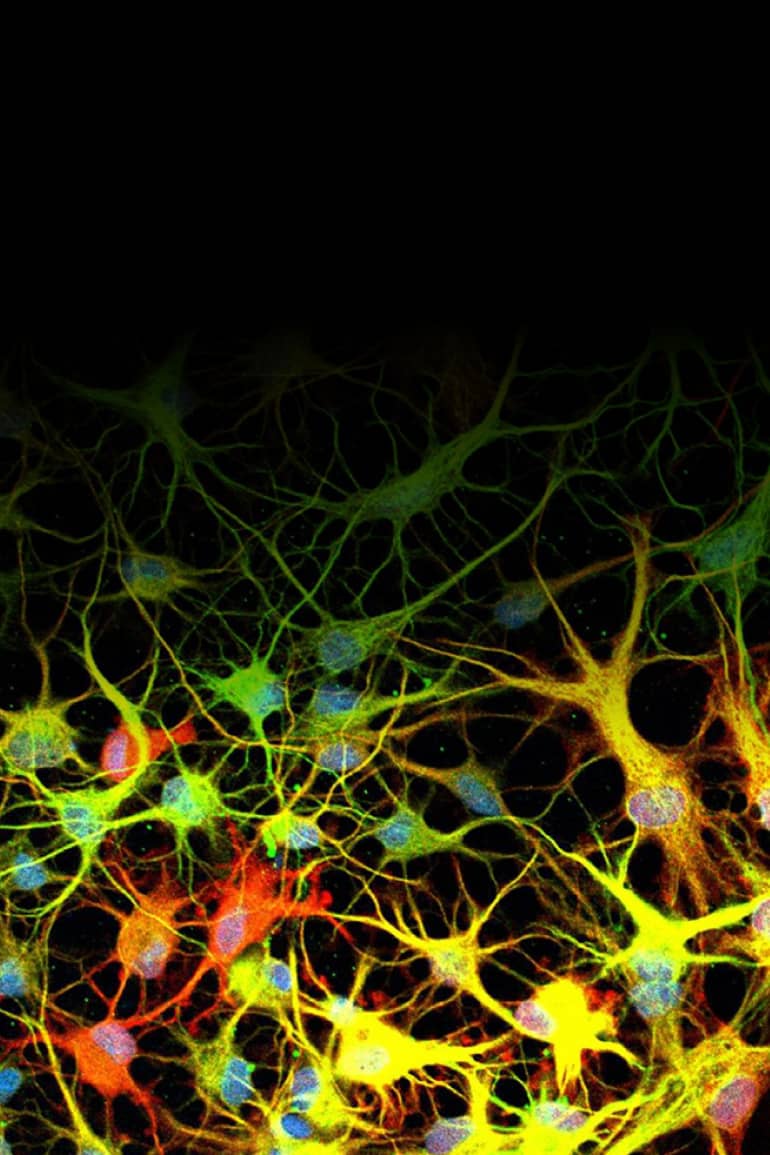
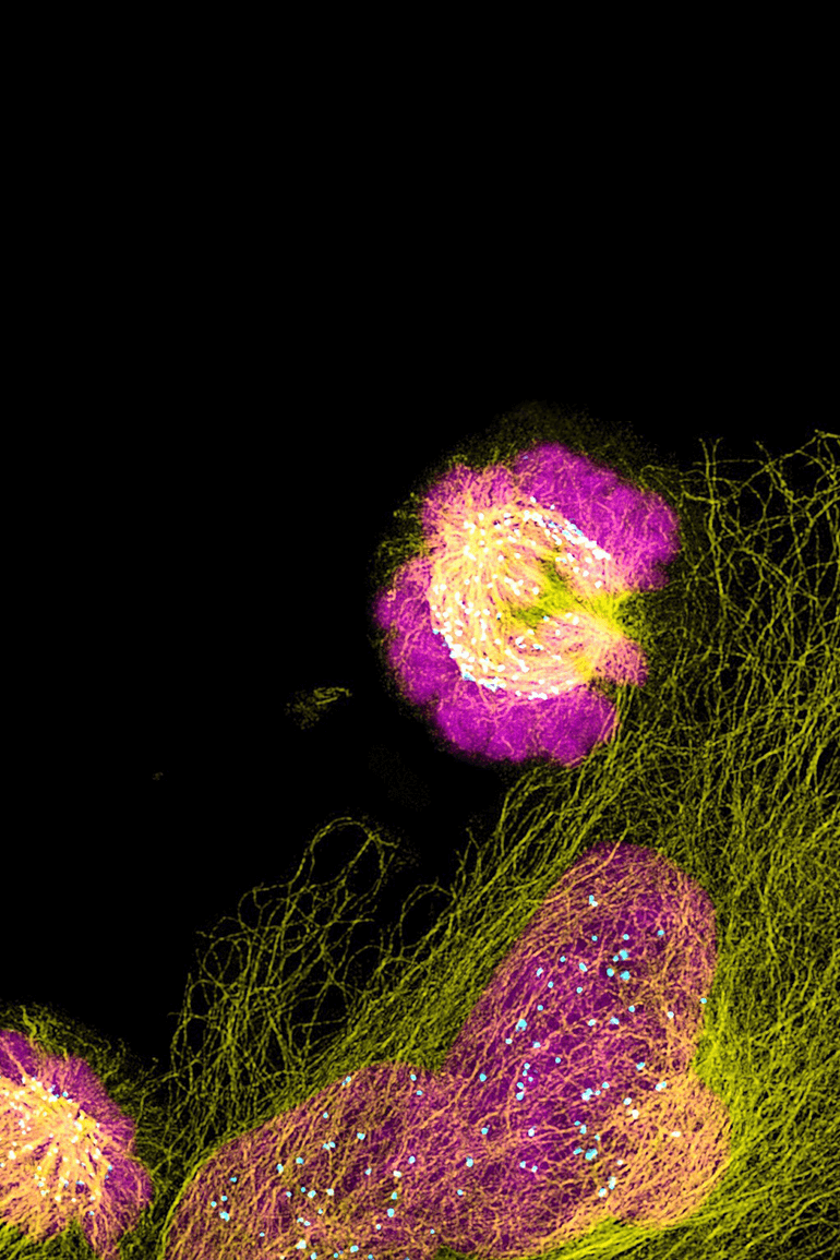
A forest of nerve cells (axons, dendrites, and dendritic spines of neurons) in the brain. Photo by Gao, Asano, Upadhyayula et al, Science 2019.
A forest of nerve cells (axons, dendrites, and dendritic spines of neurons) in the brain. Photo by Gao, Asano, Upadhyayula et al, Science 2019.
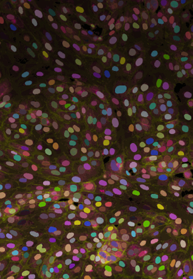
These nuclear proteins (histones) in a living roundworm embryo were imaged using a dual-view inverted selective plane illumination microscopy (diSPIM). Maximum intensity projection images are rendered in different colors for visualization. Photo provided by Abhishek Kumar, Marine Biological Laboratory, Eugene Bell Center for Regenerative Biology and Tissue Engineering.
These nuclear proteins (histones) in a living roundworm embryo were imaged using a dual-view inverted selective plane illumination microscopy (diSPIM). Maximum intensity projection images are rendered in different colors for visualization. Photo provided by Abhishek Kumar, Marine Biological Laboratory, Eugene Bell Center for Regenerative Biology and Tissue Engineering.
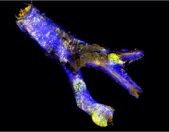
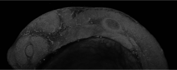
The carotid artery of a mouse with atherosclerosis. Red, yellow, and green immune cells take up cholesterol and help regulate inflammation in the arterial wall (blue). Photo by Sara McArdle, La Jolla Institute for Immunology, LJI Microscopy Core.
The carotid artery of a mouse with atherosclerosis. Red, yellow, and green immune cells take up cholesterol and help regulate inflammation in the arterial wall (blue). Photo by Sara McArdle, La Jolla Institute for Immunology, LJI Microscopy Core.
A zebrafish embryo at one day old, imaged in the Tissue Microscopy Laboratory at Texas A&M University. Photo provided by Holly Gibbs, Texas A&M University, Microscopy and Imaging Center.
A zebrafish embryo at one day old, imaged in the Tissue Microscopy Laboratory at Texas A&M University. Photo provided by Holly Gibbs, Texas A&M University, Microscopy and Imaging Center.
Super-resolution light microscopy allows scientists to view the details of subcellular organelles — units in cells with specialized functions — in living cells. Photo provided by Aaron Taylor, University of Michigan, BRCF Microscopy Core.
A two-day-old zebrafish heart viewed through a microscope. The heart muscle membrane is shown in blue and its nuclei in red. Photo provided by CZI imaging scientist Michael Weber of the Morgridge Institute for Research, in affiliation with the University of Wisconsin-Madison.
A forest of nerve cells (axons, dendrites, and dendritic spines of neurons) in the brain. Photo by Gao, Asano, Upadhyayula et al, Science 2019.
These nuclear proteins (histones) in a living roundworm embryo were imaged using a dual-view inverted selective plane illumination microscopy (diSPIM). Maximum intensity projection images are rendered in different colors for visualization. Photo provided by Abhishek Kumar, Marine Biological Laboratory, Eugene Bell Center for Regenerative Biology and Tissue Engineering.
The carotid artery of a mouse with atherosclerosis. Red, yellow, and green immune cells take up cholesterol and help regulate inflammation in the arterial wall (blue). Photo by Sara McArdle, La Jolla Institute for Immunology, LJI Microscopy Core.
A zebrafish embryo at one day old, imaged in the Tissue Microscopy Laboratory at Texas A&M University. Photo provided by Holly Gibbs, Texas A&M University, Microscopy and Imaging Center.
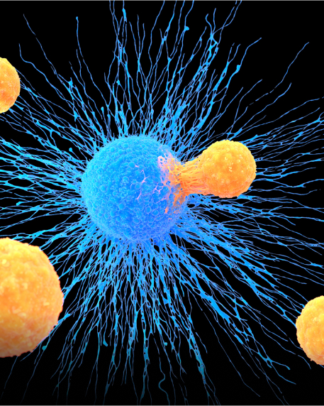
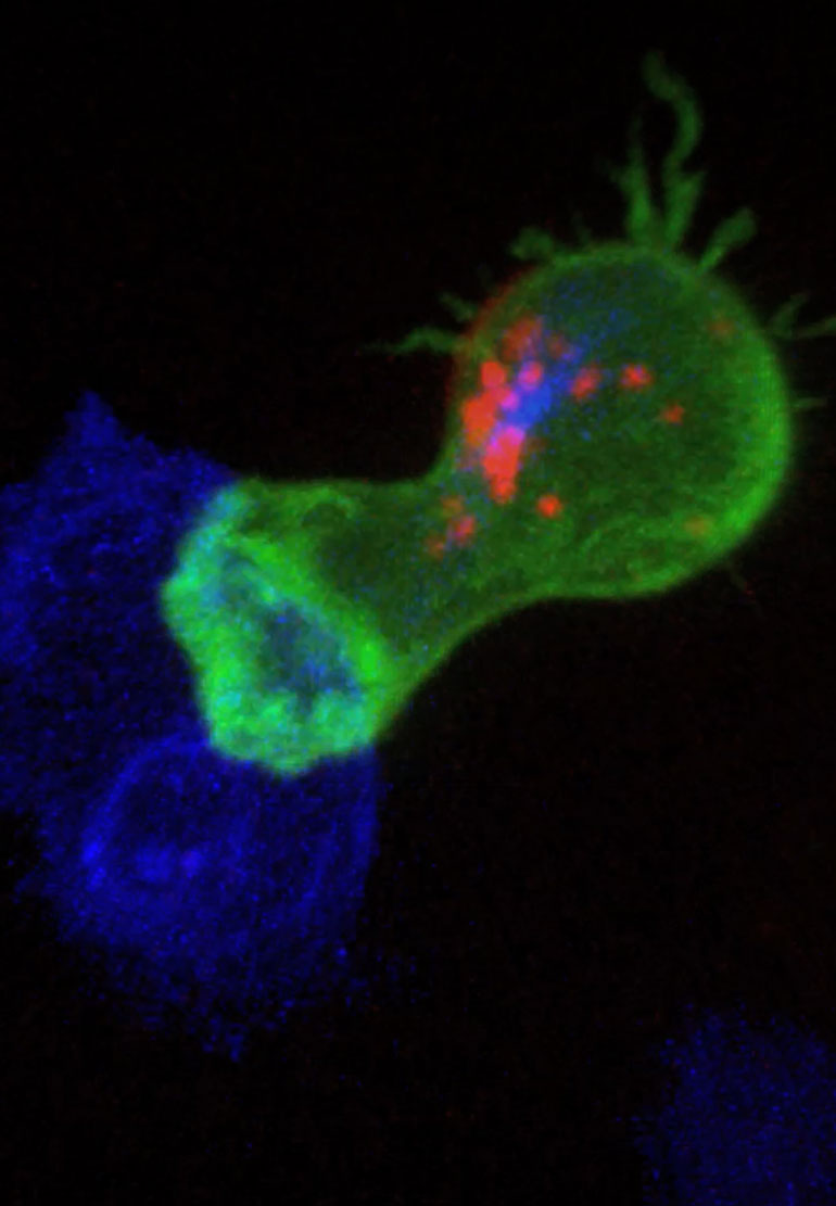
In this illustration, T lymphocytes (orange), a type of white blood cell, attack a cancer cell (blue).
In this illustration, T lymphocytes (orange), a type of white blood cell, attack a cancer cell (blue).
Frontiers of Imaging
What if researchers could view how drugs alter the behavior of cancer cells and invade immune cells deep in the body? Or visualize how viral or bacterial pathogens hijack normal cellular functions to reproduce themselves? Such insights would open up new avenues for developing treatments and cures for many diseases.
Although there have been significant advances in biomedical imaging, we are far from the ultimate goal: to observe cells and subcellular processes in living organisms and in a minimally invasive manner. CZI’s Frontiers of Imaging effort aims to accelerate the development of disruptive imaging technologies that connect biological scales—such as proteins to cells and cells to organisms. This will allow researchers to directly visualize biological processes at the necessary resolution and in context to obtain a mechanistic understanding of health and disease.
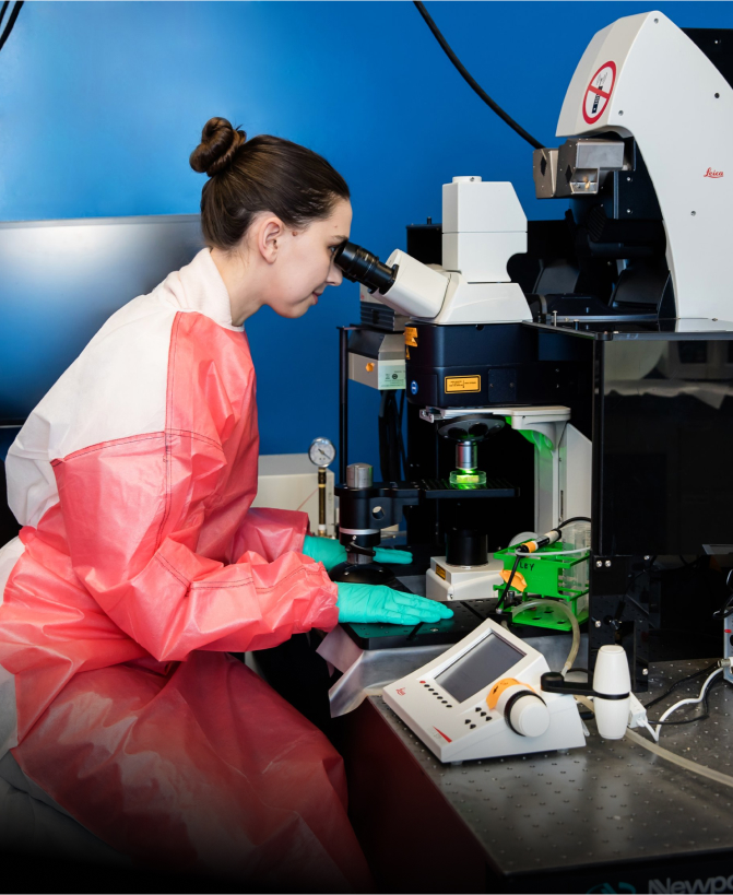
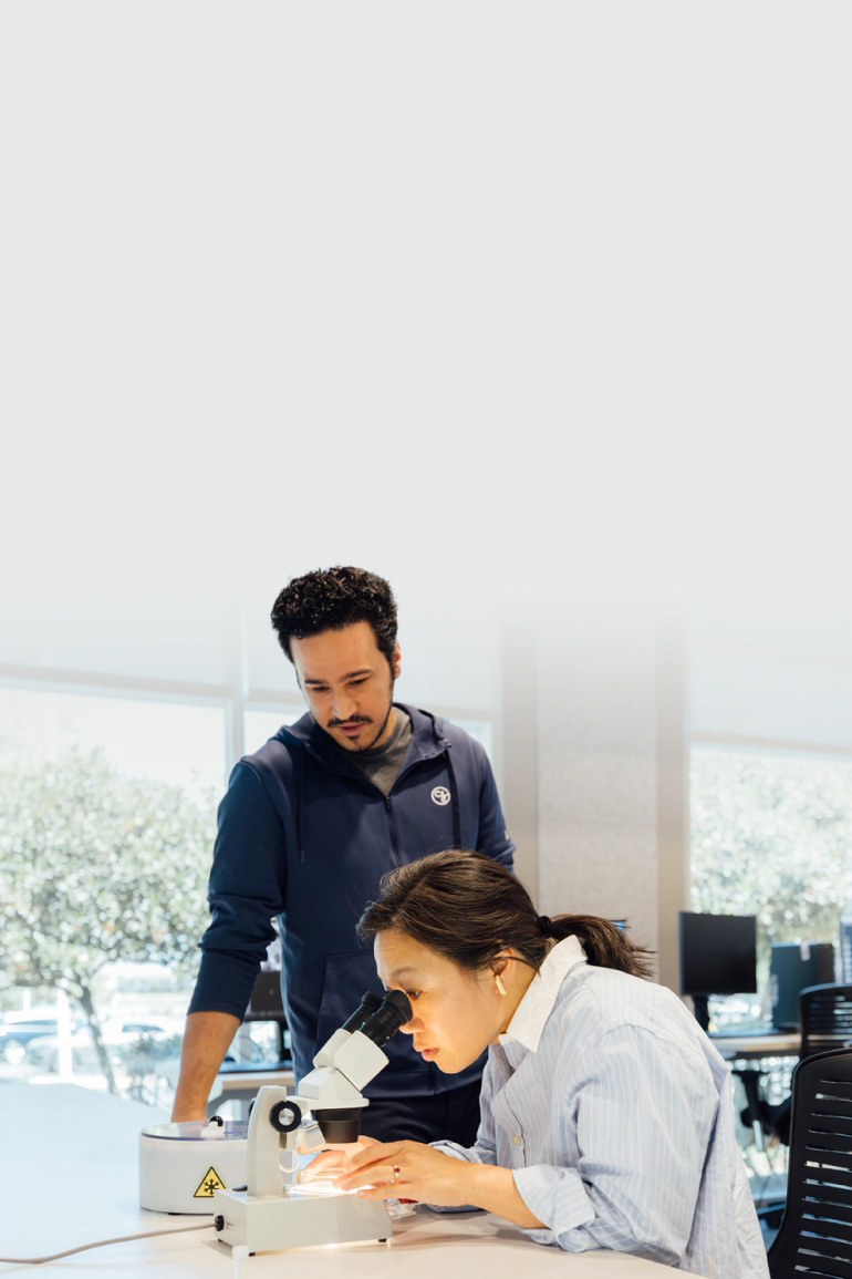
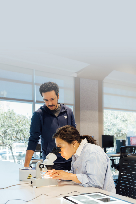
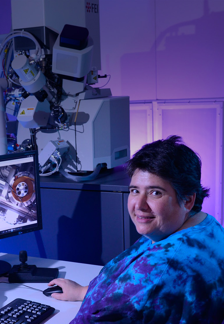
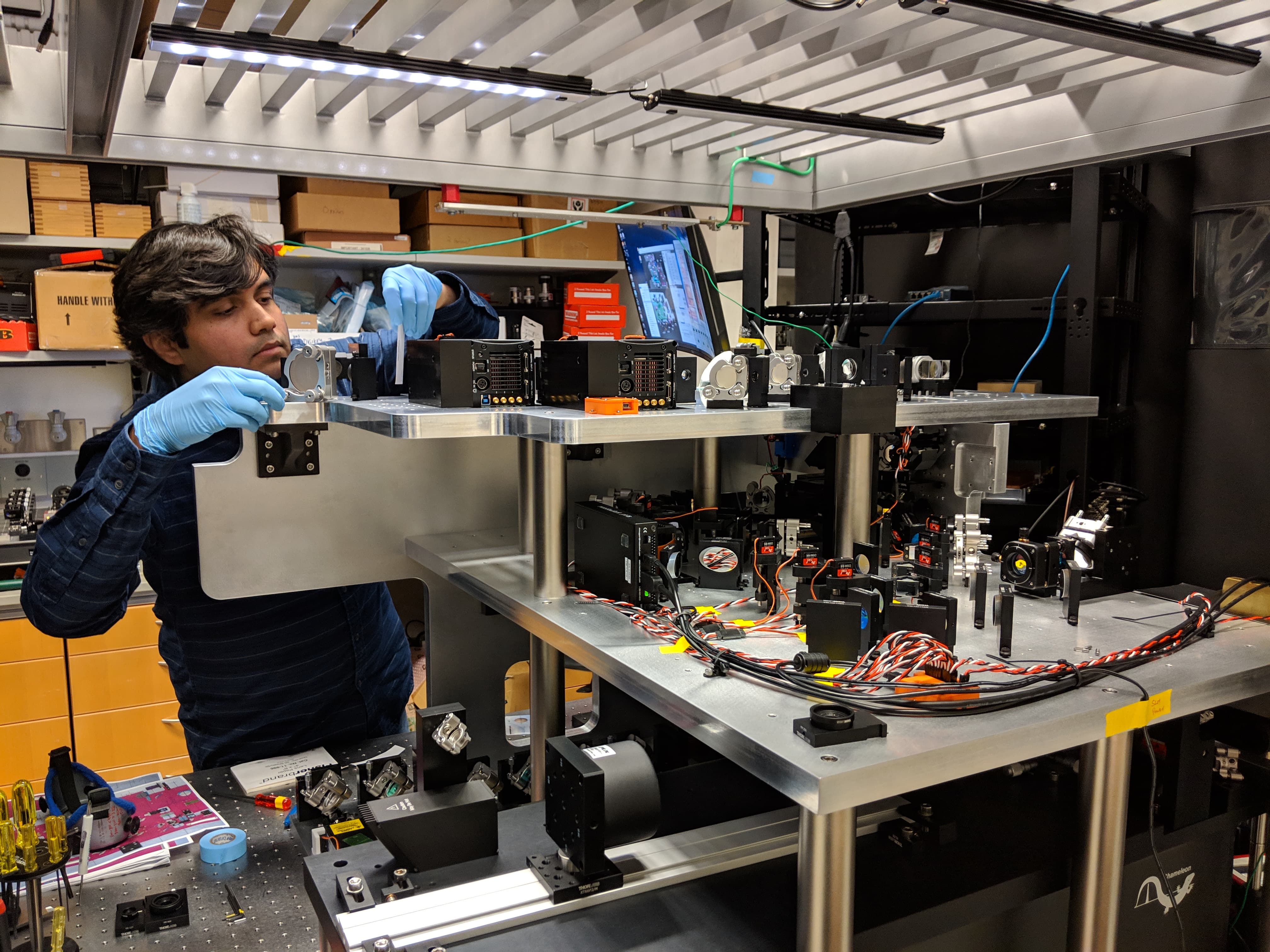
CZI imaging scientist Sara McArdle of the La Jolla Institute for Immunology looks into a microscope. Protective gowns are required, as this microscope is used in studies of infectious agents, such as Zika and Dengue viruses, in addition to a range of inflammatory disorders. Photo provided by Sara McArdle.
CZI imaging scientist Sara McArdle of the La Jolla Institute for Immunology looks into a microscope. Protective gowns are required, as this microscope is used in studies of infectious agents, such as Zika and Dengue viruses, in addition to a range of inflammatory disorders. Photo provided by Sara McArdle.
Creating Community & Building Capacity
Innovations in imaging science — new technologies, imaging modalities, and data analysis approaches — are becoming increasingly important in advancing biomedical research. To accelerate the promotion, dissemination, and efficient use of new methods and tools, CZI is supporting scientists who operate imaging facilities, which serve as hubs of imaging expertise for local communities of biomedical researchers. CZI is also supporting imaging software fellows who develop and maintain three critical imaging tools: scikit-image, FIJI / ImageJ, and CellProfiler.
On a larger scale, fostering international communities of imaging experts, scientists, imaging facility operators, and policymakers is vital to address current scientific, technical, and data challenges. To facilitate these activities, CZI supports Global BioImaging (GBI), the international network of cutting-edge imaging facilities and communities, and GBI member BioImaging North America, which brings together imaging communities spanning the United States, Canada, and Mexico. In addition, CZI is expanding access to imaging expertise, technologies, and capacity building for researchers and organizations in Africa, Latin America and the Caribbean, and former Soviet countries.
This grant program aims to drive development of complete, general-purpose, intracellular imaging probes for cryo-correlative light and electron microscopy (cryo-CLEM).
Closed
Request for Applications
Imaging
Deep Tissue Imaging (Phases 1-2)
This grant program aims to drive development of innovative imaging technologies that will advance the field of deep tissue imaging, allowing researchers to observe cells and subcellular processes at high resolution in complex tissue and through skin and bone.
This RFA supports new and existing collaborative projects to reduce imaging ecosystem fragmentation and accelerate the spread and adoption of technologies, methods, or training resources.
Closed
Request for Applications
Imaging
Dynamic Imaging
This RFA aims to advance technology directed at real-time visualization of biological processes at the level of cells and molecules.
Closed
Request for Applications
Imaging
napari Plugin Grants
This grant program will support software plugin development projects for the napari image analysis platform, a community-built, open source, and interactive tool for Python designed for browsing, annotating, and analyzing large multi-dimensional images.
This grant program supports imaging science capacity building, fellowships, and community development for imaging core facilities and nonprofit organizations in Africa, Latin America and the Caribbean, and former Soviet countries.
Closed
Request for Applications
Imaging
Visual Proteomics Imaging
This RFA supports challenge grants in visual proteomics, with the goal of advancing technology that will enable researchers to view protein molecules and their interactions inside cells at near-atomic resolution.
Closed
Request for Applications
Imaging
Imaging Scientists Cycles 1-2
This RFA supports CZI Imaging Scientists at imaging centers worldwide who will leverage biology, microscopy hardware, and imaging software to accelerate biomedicine and improve the imaging tools scientists use.
Imaging Scientists Cycles 1-2
- $17.8 Million
- $10.2 Million
News & Stories
Interested in learning more about our work in imaging? Get the latest information from the links below.
We use cookies to help us improve the site and to inform our marketing and digital content efforts. If you choose ‘Don’t Enable,’ sites you’re logged into – like Facebook and Twitter – may still be able to identify you as a visitor to this site. Learn more.


 Contact the Imaging team